Welcome to
India's Largest Medical Learning Platform
Trusted by 16,50,000+ HCPs to advance their clinical practice through LIVE CME, expert-led courses, and evidence-based medical education.
MedWiki®Experiential medicine at your fingertips
A collaborative medical knowledge hub built from expert discussions, case studies, and clinical insights shared by doctors across specialties
Clinical VideoWatch expert-led clinical discussions
Expert-led clinical videos designed to deliver practical insights, treatment strategies, and real-world case learning across multiple medical specialties.
LiveCMEReal-time collaboration could not have been easier
Engage in live Continuing Medical Education sessions led by leading specialists, featuring expert discussions on the latest medical guidelines and practices.
AgentIntelligent clinical assistance at your fingertips
Intelligent AI assistants that help doctors quickly access medical knowledge, clinical insights, and relevant resources to support faster and smarter decision-making.
SPQKnow the Doctor's Voice
Interactive polls and quizzes that allow doctors to test their knowledge and gain insights into real-world clinical opinions from the medical community.
You’re viewing a preview of this page - Log in to explore full content and personalized features.
Chat with Our ExpertsPublic Beta
Start with an Agent or ask freely


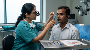
Paralytic and restrictive strabismus: surgical planning
Strabismus is a disorder of ocular alignment that can impair binocular vision and stereopsis and may cause diplopia and psychosocial difficulties. Paralytic and restrictive strabismus are important forms of ocular misalignment that require different approaches to surgical planning. Paralytic strabismus results from weakness or loss of function of an extraocular muscle, commonly due to cranial nerve dysfunction, whereas restrictive strabismus arises from mechanical limitation of eye movement caused by factors such as fibrosis, inflammation, scarring, or abnormal tissue attachments. Surgical management aims to improve ocular alignment, reduce diplopia and abnormal head posture, and optimise binocular function where feasible. Accurate diagnosis, assessment of ocular motility and deviation, identification of the underlying cause, and confirmation of disease stability are essential for selecting an appropriate surgical strategy and achieving realistic outcomes.[1]Preoperative assessmentEstablish the cause and pattern: Assess the onset, progression, ocular motility, deviation in different gaze positions, and relevant neurologic, orbital, traumatic, or surgical historyQuantify ocular deviation: Perform prism-cover testing at distance and near, document deviations in primary and diagnostic gaze positions, and assess torsion when indicatedEvaluate muscle function: Determine whether the affected muscle has reduced force generation, whether an opposing muscle is contracted, and whether mechanical restriction limits passive movementAssess binocular function: Document diplopia, suppression, stereopsis, compensatory head posture, and the patient's functional prioritiesInvestigate associated disease: Obtain appropriate neurologic or orbital evaluation when the presentation suggests cranial nerve palsy, orbital inflammation, thyroid eye disease, trauma, or a space-occupying lesionSurgical planning in paralytic strabismusConfirm stability: When clinically appropriate, defer definitive surgery until the deviation and underlying condition have stabilized, because spontaneous recovery may occur in some acquired cranial nerve palsiesAddress deficient muscle force: Select procedures according to the affected nerve and muscle, deviation magnitude, residual muscle function, and pattern of incomitanceConsider transposition procedures: In selected cases of severe muscle paresis, transposition of functioning extraocular muscles may improve alignment when direct action of the paretic muscle is inadequateManage antagonist contracture: Recession of a tight antagonist may be required when contracture contributes substantially to the deviation, but the procedure must account for residual muscle function and postoperative alignmentPlan for adjustable sutures selectively: Adjustable techniques may be considered in appropriate patients when postoperative refinement of alignment could be beneficialSurgical planning in restrictive strabismusIdentify the mechanical cause: Determine whether restriction arises from fibrosis, scarring, inflammation, trauma, thyroid eye disease, or an abnormal muscle pathPerform forced-duction testing: Passive restriction should be assessed when clinically indicated, often under anesthesia, to help distinguish mechanical limitation from weaknessRelieve restriction when appropriate: Recession of the restricted muscle or release of restrictive tissue may improve movement, depending on the cause and anatomical findingsTreat the underlying disorder: Active orbital inflammation or unstable thyroid eye disease may require medical management and stabilization before elective alignment surgeryAnticipate residual incomitance: Surgery may improve primary-position alignment without restoring normal movement in every direction of gazeFactors influencing procedure selectionClinical findingSurgical planning implicationSevere paresis with inadequate residual muscle actionConsider whether a transposition procedure is appropriate after assessing the risk of anterior segment ischemia.Positive forced-duction testInvestigate and address mechanical restriction rather than treating the deviation as isolated muscle weakness.Multiple rectus muscles requiring surgeryEvaluate anterior segment circulation and consider the cumulative vascular risk of operating on several rectus muscles.Significant vertical deviation or torsionIncorporate vertical alignment and torsional symptoms into procedure selection and counseling.Postoperative considerationsAssess alignment, ocular motility, diplopia, and head posture after surgery, recognizing that residual or recurrent deviation may require further treatmentCounsel patients that improved primary-position alignment does not guarantee restoration of full ocular motility or binocular single visionConsider staged surgery or additional interventions when the deviation is complex, the underlying disease remains active, or the initial result is insufficientConclusionOculomotor nerve palsy presents a particular challenge in strabismus management because it can affect four of the six extraocular muscles, resulting in complex ocular misalignment. Associated features, including ptosis, pupillary dysfunction, amblyopia, oculomotor synkinesis, impaired Bell’s phenomenon, superior oblique overaction, and lateral rectus contracture, may further complicate treatment and surgical planning. Successful management of paralytic and restrictive strabismus depends on accurately distinguishing muscle weakness from mechanical restriction and tailoring intervention to the underlying pathology. Comprehensive ocular motility assessment, appropriate investigations, confirmation of disease stability, and realistic preoperative counselling are essential for selecting the appropriate procedure. The primary goal is to improve ocular alignment, reduce diplopia where possible, and enhance visual function, recognising that full restoration of ocular motility may not always be achievable.[2]Reference(s):1.Martínez-Abad A, Siverio-Colomina A, Amesty MA, Díez-de-la-Uz R, Cantó-Cerdán M. Comparison of the Surgical Treatment for Strabismus According to Its Type: Esotropia Versus Exotropia. J Clin Med. 2026;15(2):795. Published 2026 Jan 19. doi:10.3390/jcm15020795. https://pmc.ncbi.nlm.nih.gov/articles/PMC12841767/2.Rifqi K, Loebis R. Combined recession-resection in the management of paralytic strabismus: simplification of the complex (a case report). Pan Afr Med J. 2024;48:165. Published 2024 Aug 9. doi:10.11604/pamj.2024.48.165.44739. https://pmc.ncbi.nlm.nih.gov/articles/PMC11614114/
Case-Based Discussion on Guillain-Barré Syndrome


Dr. Voloju Adithya
Laparoscopic Colorectal Cancer Surgery: Evidence and Challenges
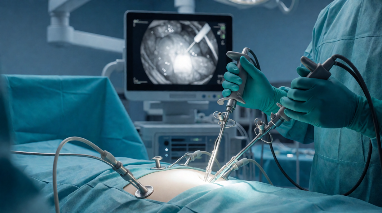

Dr. Kshitij Arun Manerikar
Do you think routine prophylactic antibiotics should be avoided in acute pancreatitis without suspected or confirmed infection?
Do you think routine prophylactic antibiotics should be avoided in acute pancreatitis without suspected or confirmed infection?
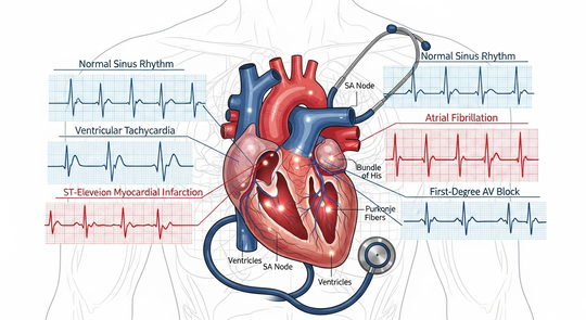
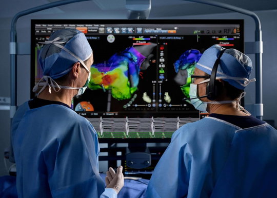
The Arrhythmia Playbook: From Recognition to Management
















NMC Releases Detailed Stipend List for All Indian Medical Colleges
This document is compilation of the National Medical Commission (NMC) stipend details reported by 804 medical colleges across India, covering undergraduate (UG/MBBS interns), postgraduate (PG), and senior resident (SS) trainees. Reports were received between 2024 and January 2026. The data captures: whether stipend reports were submitted to NMC, state government stipend amounts, college-provided stipend amounts, affiliating universities, and institution type (Government / Private / Trust / Society). Several colleges were newly established (batch started 2023–2025) with no interns yet, while a few colleges were closed or had courses unavailable. Colleges which did not provide the said stipend information or refused to share data have been explicitly flagged.
A Clinical Approach to DSPN & DAN


Prof. Dr. Vitull K. Gupta

Whole-body MRI and PET-CT in multiple myeloma and bone metastases: staging and treatment response
Multiple myeloma (MM) is a haematological malignancy characterised by the clonal proliferation of plasma cells in the bone marrow and the production of monoclonal immunoglobulins or their components. Bone disease is a major feature of MM, while other clinical manifestations include anaemia, hypercalcaemia, and renal impairment. Imaging plays a central role in disease detection, initial staging, assessment of disease extent, and evaluation of treatment response. Whole-body magnetic resonance imaging (MRI) and positron emission tomography–computed tomography (PET-CT) provide complementary information for assessing MM and bone metastases from other malignancies. Whole-body MRI is highly sensitive to bone marrow infiltration, including focal and diffuse disease that may precede visible cortical destruction. PET-CT combines metabolic and anatomical information to identify metabolically active lesions, detect extramedullary involvement, and monitor metabolic response to treatment. The choice of imaging modality depends on the suspected or established malignancy, the clinical question, baseline findings, and the treatment setting. Understanding the strengths and limitations of these techniques is essential for accurate staging and consistent assessment of treatment response.[1]Whole-body MRI for staging and disease assessmentDetect marrow infiltration: Whole-body MRI, particularly diffusion-weighted imaging, can identify focal and diffuse marrow abnormalities before structural bone destruction becomes apparentAssess myeloma burden: MRI helps identify the distribution and number of focal lesions, supporting the evaluation of suspected myeloma and selected patients with smoldering diseaseEvaluate complications: Targeted MRI is important when spinal cord compression, nerve involvement, or other soft-tissue complications are suspectedInterpret response cautiously: Changes in marrow signal and diffusion characteristics may persist during treatment or recovery, so imaging findings should be interpreted alongside clinical and laboratory evidenceUse standardized reporting: The Myeloma Response Assessment and Diagnosis System (MY-RADS) supports consistent whole-body MRI acquisition, interpretation, and response assessmentFig: MRI illustration of a diffuse medullary infiltration pattern in myeloma using T1w (a) and STIR (b) sequences. It is characterized by decreased signal intensity of the bone marrow on T1w sequences (a) and a correspondingly increased signal on STIR (b) sequences. On T1w images (a), the signal intensity of the bone marrow is equal to the intervertebral disc and the muscle, which is known as the “bright disc” sign[2]PET-CT for staging and treatment responseIdentify metabolically active disease: Fluorine-18 fluorodeoxyglucose (FDG) PET-CT detects glucose-avid lesions and provides information about disease activity alongside CT findingsEvaluate extramedullary involvement: PET-CT can identify metabolically active soft-tissue disease outside the bone marrow and help establish disease distributionAssess treatment response: In multiple myeloma, PET-CT can demonstrate reduced or absent metabolic activity following treatment, including when residual structural abnormalities remainRecognize limitations: False-negative findings can occur in FDG-nonavid disease, while inflammation, infection, and treatment-related changes can produce false-positive uptakeMaintain comparability: When serial imaging is indicated, comparison with baseline studies using a consistent modality and standardized interpretation criteria improves assessment of response or progressionImaging of bone metastasesMRI is particularly useful for suspected spinal or epidural involvement, marrow disease, and assessment of possible metastatic spinal cord compressionPET-CT may help identify metabolically active skeletal lesions and additional sites of disease, depending on the primary tumor's FDG avidityCT remains important for evaluating cortical destruction, structural integrity, and fracture risk, which functional imaging alone cannot adequately establishImaging findings should be reviewed alongside symptoms, examination findings, primary tumor characteristics, and other investigations to guide multidisciplinary managementSelecting imaging for the clinical questionClinical considerationImplication for imaging selectionSuspected myeloma with negative or inconclusive initial imagingAdditional whole-body imaging may be required because different modalities detect different patterns of disease.Suspected impending pathological fractureDedicated CT or radiographs may be needed to assess cortical integrity and structural stability.Lesion with uncertain metabolic activityCorrelation with anatomic imaging, clinical findings, and interval assessment may help clarify significance.Discordant imaging and laboratory responseMultidisciplinary review is appropriate before changing treatment based on imaging alone.ConclusionWhole-body MRI is increasingly used to evaluate disease extent across a range of malignancies, although standardised imaging protocols and reporting approaches remain important for consistent interpretation. In multiple myeloma and bone metastases, whole-body-MRI and PET-CT provide complementary information and should be selected according to the malignancy and clinical question. MRI is particularly valuable for detecting bone marrow involvement and assessing spinal and other soft-tissue complications, whereas PET-CT provides metabolic information that supports disease staging and treatment-response assessment. Consistent imaging protocols, comparison with baseline studies, and integration of imaging findings with clinical and laboratory data are essential for accurate disease evaluation, treatment planning, and follow-up.[3]Reference(s):1.Mulé S, Reizine E, Blanc-Durand P, et al. Whole-Body Functional MRI and PET/MRI in Multiple Myeloma. Cancers (Basel). 2020;12(11):3155. Published 2020 Oct 27. doi:10.3390/cancers12113155. https://pmc.ncbi.nlm.nih.gov/articles/PMC7693006/2.Treitl KM, Ricke J, Baur-Melnyk A. Whole-body magnetic resonance imaging (WBMRI) versus whole-body computed tomography (WBCT) for myeloma imaging and staging. Skeletal Radiol. 2022;51(1):43-58. doi:10.1007/s00256-021-03799-4. https://pmc.ncbi.nlm.nih.gov/articles/PMC8626374/3.Parillo M, Mallio CA. The Whole-Body MRI Reporting and Data System Guidelines for Prostate Cancer (MET-RADS-P), Multiple Myeloma (MY-RADS), and Cancer Screening (ONCO-RADS). Cancers (Basel). 2025;17(2):275. Published 2025 Jan 16. doi:10.3390/cancers17020275. https://pmc.ncbi.nlm.nih.gov/articles/PMC11763526/
Evidence for Optimal Therapy in Managing IBS
Landscape Revolution in Cancer


Dr. Suresh Advani

Everything a medical professional
needs in one place
From clinical literature to peer discussions, CLIRNET is designed around how medical professionals actually learn and grow.